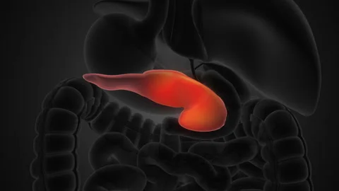
Pancreatic Cells Types Structure Functions Diseases Biology Diagrams Gross Anatomy. The pancreas is a narrow, 6-inch long gland that lies posterior and inferior to the stomach on the left side of the abdominal cavity. The pancreas extends laterally and superiorly across the abdomen from the curve of the duodenum to the spleen. The head of the pancreas, which connects to the duodenum, is the widest and most
The pancreas (plural pancreata) is an organ of the digestive system and endocrine system of vertebrates. In humans, it is located in the abdomen behind the stomach and functions as a gland. The pancreas is a mixed or heterocrine gland, i.e., it has both an endocrine and a digestive exocrine function. [2] 99% of the pancreas is exocrine and 1%
Anatomy, Abdomen and Pelvis, Pancreas Biology Diagrams
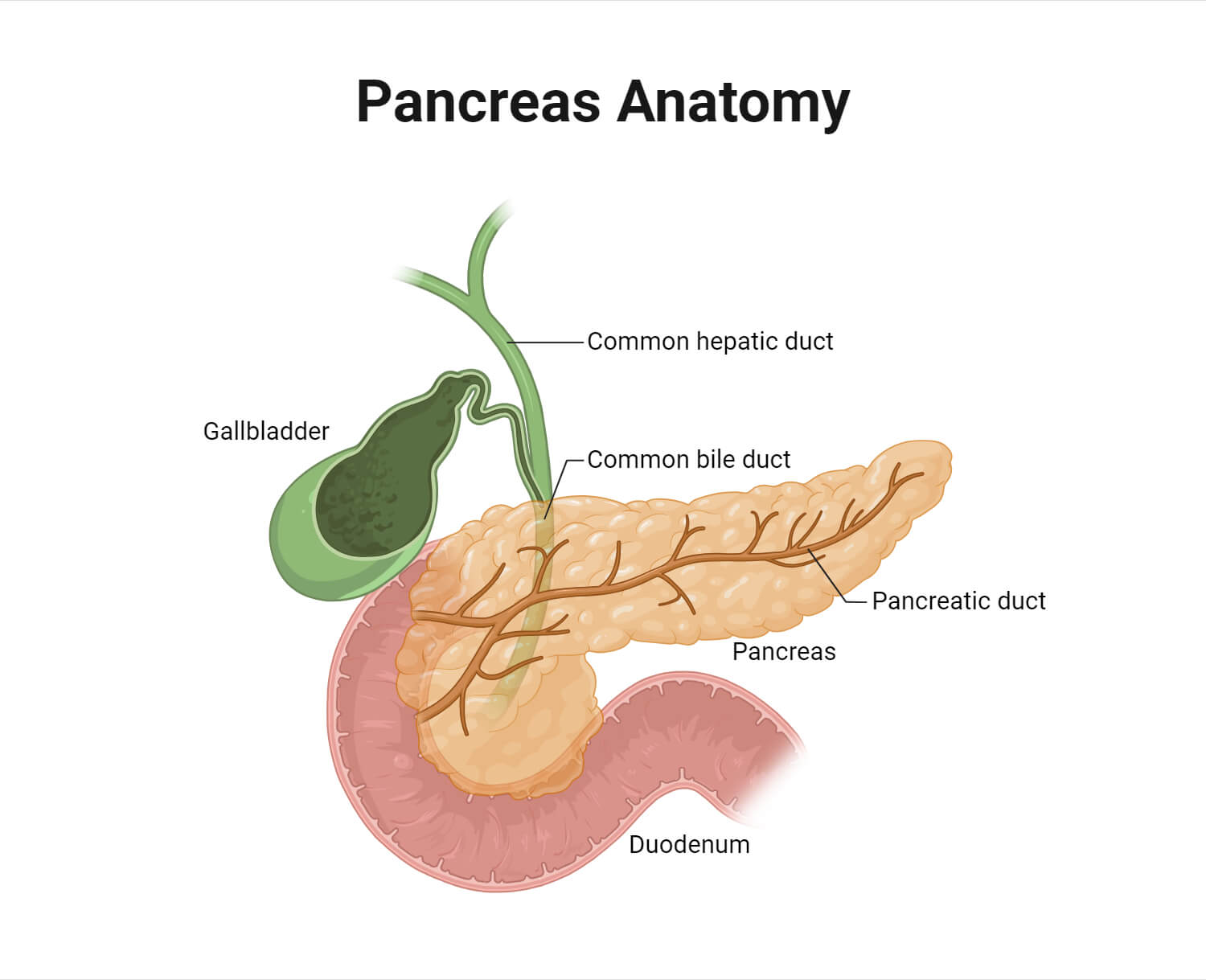
Pancreas anatomy. Parts of the pancreas include the: Head: The wider part of your pancreas that sits in the curve of your duodenum. On average, a healthy human pancreas weighs around 91.8 grams (0.20 pounds). That's about the same as a deck of playing cards. Conditions and Disorders. Pancreas Anatomy. The pancreas measures about 20 centimeters in length (about 8 inches) and weighs between 75 to 90 grams (heavier in men). The bulk of the pancreatic tissue is dedicated to its exocrine function, while only 1% to 2% of the pancreas is responsible for the endocrine component. Human Intestinal Parasites - Causes, Symptoms The Stomach, Gallbladder, and Pancreas Explore the anatomy and roles of the stomach, gallbladder, and pancreas with Innerbody's interactive 3D model. by Tim Taylor Last The stomach, gallbladder, and pancreas are three of the most important digestive organs in the human body. These organs work together to produce and store secretions that

The pancreas is an accessory organ and exocrine gland of the digestive system, as well as a hormone producing endocrine gland.It is a retroperitoneal organ consisting of five parts and an internal system of ducts. The pancreas is supplied by pancreatic arteries stemming from surrounding vessels and is innervated by the vagus nerve (CN X), celiac plexus, and superior mesenteric plexus.

The Pancreas: Anatomy and 3D Illustrations Biology Diagrams
The pancreas is an extended, accessory digestive gland that is found retroperitoneally, crossing the bodies of the L1 and L2 vertebrae on the posterior abdominal wall. The pancreas lies transversely in the upper abdomen between the duodenum on the right and the spleen on the left. It is divided into the head, neck, body, and tail. The head lies on the inferior vena cava and the renal vein and The pancreas is a vital glandular organ in the human body that serves both endocrine and exocrine functions. It plays a critical role in digestion and metabolism. [2] Structurally, it is soft, elongated, and tapered, with a lobular appearance due to its glandular tissue.The pancreas is composed of acinar cells, which produce digestive enzymes, and pancreatic islets, which regulate hormone